-
Blood pressure monitoring with plethysmograph offers the least discomfort to the subject; however, it provides only a relative indication of the well being of the circulatory system rather than providing absolute values for diastolic and systolic pressure.
-
Digital blood pressure monitors are now-a-days often used in many intensive care units. Although diastolic and systolic arterial pressure are commonly monitored, mean arterial
pressure and venous pressure are also be monitored in some instances. -
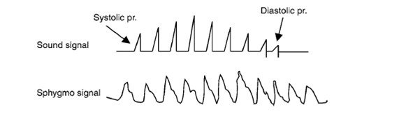
If the finger pulse pick up is also used with the subject, then it is possible to measure the diastolic pressure by noting when the signal increases and reaches a maximum and falls.
-
The signal will rise as the cuff pressure is released below systolic pressure. Then, when it reaches the lower diastolic pressure value, the artery fully admits blood flow and hence the signal increases. When the rise is maximum and then it falls, note the pressure, which is the diastolic pressure.
-
After reaching maximum value, the pulse signal remains constant as it should be, whereas the sound signal is absent after diastolic pressure. If a suitable meter is provided on the output of the finger pulse signal, using a 500 uA mini meter (such as the one available as VU-meter for
audio parts shops for Rs. 30/-), then the observation of the peak of the signal would be easier.
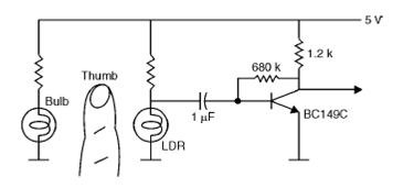
Circuit Diagram:
PRINCIPLE OF PPG
PPG makes uses of low-intensity infrared (IR) light. When light travels through biological tissues it is absorbed by bones, skin pigments and both venous and arterial blood. Since light is more strongly absorbed by blood than the surrounding tissues, the changes in blood flow can be detected by PPG sensors as changes in the intensity of light.
The voltage signal from PPG is proportional to the quantity of blood flowing through the blood vessels. Even small changes in blood volume can be detected using this method, though it cannot be used to quantify the amount of blood. A PPG signal has several components including volumetric changes in arterial blood which is associated with cardiac activity, variations in venous blood volume which modulates the PPG. Some major factors affecting the recordings from the PPG are site of measurement and the contact force between the site and the sensor.
Blood flow variations mostly occur in the arteries and not in the veins. The finger pulse pick up is one the simplest of pulse transducers to know the arterial blood flow and also to determine the heart rate. The same signal can also be used to determine the Diastolic pressure in the indirect blood pressure measurement. The circuit provides the output of 100-200mV at the collector of the transistor. This can be directly observed on a long persistence CRO or the Digital storage oscilloscope. The reason why general purpose oscilloscopes are not useful is that here, the time base is to be set as 2 seconds per sweep and the signal is coming so slowly that the trace fades away before permitting one to view the complete sphygmogram. In a Digital storage oscilloscope, since only the stored data is repeatedly presented, the trace is visible, even though the effective sweep is 2 second.
In DSOs, there is provision to send the waveform data, after “freezing” it to a PC through the
interface provided on the scope. Usually RS232 interface is simple, and the copy can be printed on the PC printer.
The data file is available for processing as well. The use of the photodiode can be similarly made as the LDR, but the current through the diode is operated at a much less value since the diode is actually worked in reverse mode.
WORKING OF INSTRUMENT
Photoplethysmography (PPG) is a simple optical technique used to detect volumetric changes in blood in peripheral circulation. It is a low cost and non-invasive method that makes measurements at the surface of the skin.
The technique provides valuable information related to our cardiovascular system. Recent advances in technology has revived interest in this technique, which is widely used in clinical physiological measurement and monitoring.
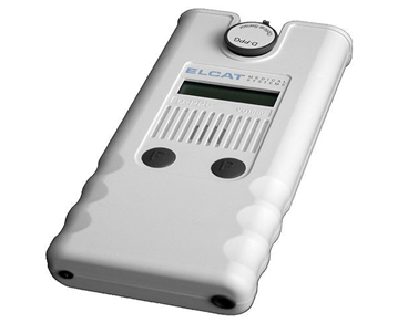
The Vasoquant 1000 is a 1-channel-photoplethysmograph that is used for venous function diagnosis.
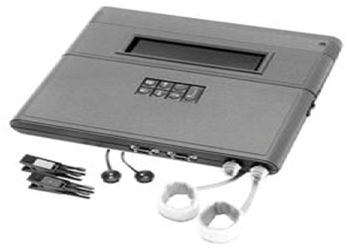
The Vasoquant 2000 is a 2 channeled photoplethysmograph provides diagnosis for venous and acral arterial functions.